Here’s a detailed overview of an X-ray of the knee:
🔹 What a Knee X-Ray Is
- A radiographic image of the knee joint
- Shows bones: femur (thigh), tibia (shin), patella (kneecap)
- Can detect:
- Fractures or bone breaks
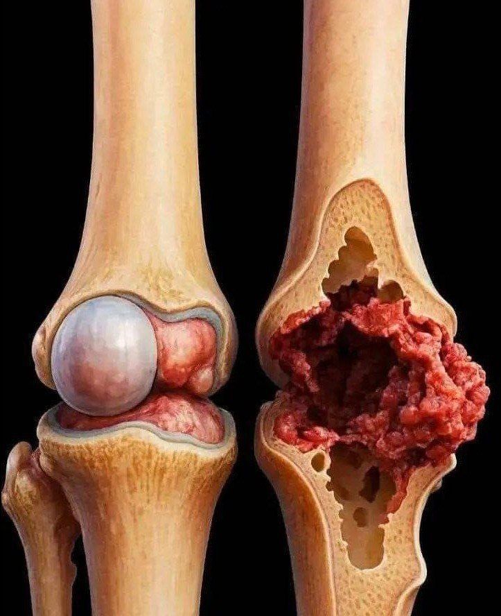
- Arthritis or joint space narrowing
- Bone tumors or infections
- Alignment issues
🔹 Types of Knee X-Rays
- AP (Anteroposterior) view – front-to-back of knee
- Lateral view – side view of knee
- Sunrise/skyline view – patella (kneecap) view
- Weight-bearing view – standing, to assess joint space and arthritis
🔹 How It’s Done
- You sit or lie on the X-ray table
- The technician positions your knee for the required views
- Minimal radiation exposure (very low risk)
- Takes 1–5 minutes per view
🔹 Preparation
- Usually no special prep needed
- Remove metal objects (rings, jewelry, knee braces)
- Tell technician if pregnant
🔹 What Can Be Seen
- Fractures or cracks in bones
- Osteoarthritis – narrowed joint space, bone spurs
- Dislocations – misalignment of patella or joint
- Bone tumors or cysts (rarely)
- Signs of previous surgery (pins, plates, screws)
⚠️ Limitations
- Does not show soft tissue well (ligaments, tendons, cartilage)
- For ligament injuries, meniscus tears, or cartilage damage → MRI is preferred
🧠 Quick Summary
- X-ray = best for bones
- Quick, painless, minimal radiation
- Essential first step in diagnosing knee pain, injury, or arthritis
If you want, I can make a simple labeled diagram showing a knee X-ray with bones and common injuries marked—great for easy understanding. Do you want me to do that?
