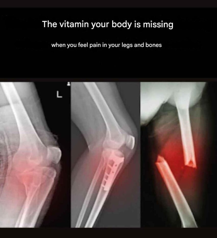
Here’s a detailed overview of X-ray imaging for a proximal tibia fracture and post-fixation assessment:
1. Anatomy Overview
- The proximal tibia is the upper part of the shinbone, just below the knee joint.
- Key structures seen on X-ray:
- Tibial plateau (medial and lateral)
- Epiphysis and metaphysis
- Adjacent fibula head
- Knee joint space
2. X-ray Views for Proximal Tibia Fractures
- Anteroposterior (AP) View – Shows width of the tibial plateau and fracture lines.
- Lateral View – Shows sagittal plane alignment, posterior slope, and displacement.
- Oblique Views (optional) – Help visualize complex fracture patterns.
3. Types of Proximal Tibia Fractures
- Tibial plateau fractures: Can involve lateral, medial, or both plateaus.
- Extra-articular fractures: Fracture below the plateau, not entering joint space.
- Comminuted fractures: Bone broken into multiple fragments.
Key X-ray findings may include:
- Displacement of bone fragments
- Depression of tibial plateau
- Intra-articular extension
4. Post-Fixation (Post-Op) X-ray Assessment
After surgical fixation (ORIF – Open Reduction Internal Fixation):
Check for:
- Proper alignment – Tibial plateau should be level and aligned with femoral condyles.
- Hardware placement – Screws, plates, or rods should follow planned positions.
- Joint space integrity – No collapse or depression into the knee joint.
- Bone healing – Early signs of callus formation may appear in follow-up X-rays.
Common Post-Fixation Complications on X-ray
- Malalignment or step-off of tibial plateau
- Screw or plate loosening
- Non-union or delayed union
- Secondary fracture around hardware
5. Follow-Up
- X-rays are typically done immediately post-op, then at 2–6 weeks, and periodically until fracture heals.
- Physical therapy is guided based on fracture type, fixation stability, and X-ray findings.
💡 Tip: Always compare pre-op and post-op X-rays to assess fracture reduction and hardware placement. Accurate imaging helps prevent long-term knee dysfunction.
If you want, I can create a visual guide showing a proximal tibia fracture X-ray, before and after fixation, highlighting key features and hardware placement.
Do you want me to make that?
