Human heart anatomy diagram
A human heart anatomy diagram visually represents the structure of the heart and how blood flows through its chambers, valves, and vessels. Understanding this is crucial for learning circulatory function, heart health, and disease mechanisms.
🔹 Key Parts of the Heart
- Chambers
- Right atrium (RA): Receives deoxygenated blood from the body via the superior and inferior vena cava.
- Right ventricle (RV): Pumps blood to the lungs through the pulmonary artery.
- Left atrium (LA): Receives oxygenated blood from the lungs via pulmonary veins.
- Left ventricle (LV): Pumps oxygen-rich blood to the entire body via the aorta.
- Valves
- Tricuspid valve: Between right atrium and ventricle; prevents backflow.
- Pulmonary valve: Between right ventricle and pulmonary artery.
- Mitral (bicuspid) valve: Between left atrium and ventricle.
- Aortic valve: Between left ventricle and aorta.
- Major Vessels
- Aorta: Main artery carrying oxygenated blood to the body.
- Superior and inferior vena cava: Return deoxygenated blood from the body.
- Pulmonary arteries: Carry deoxygenated blood to the lungs.
- Pulmonary veins: Carry oxygenated blood from the lungs.
- Other Structures
- Septum: Muscular wall separating left and right sides.
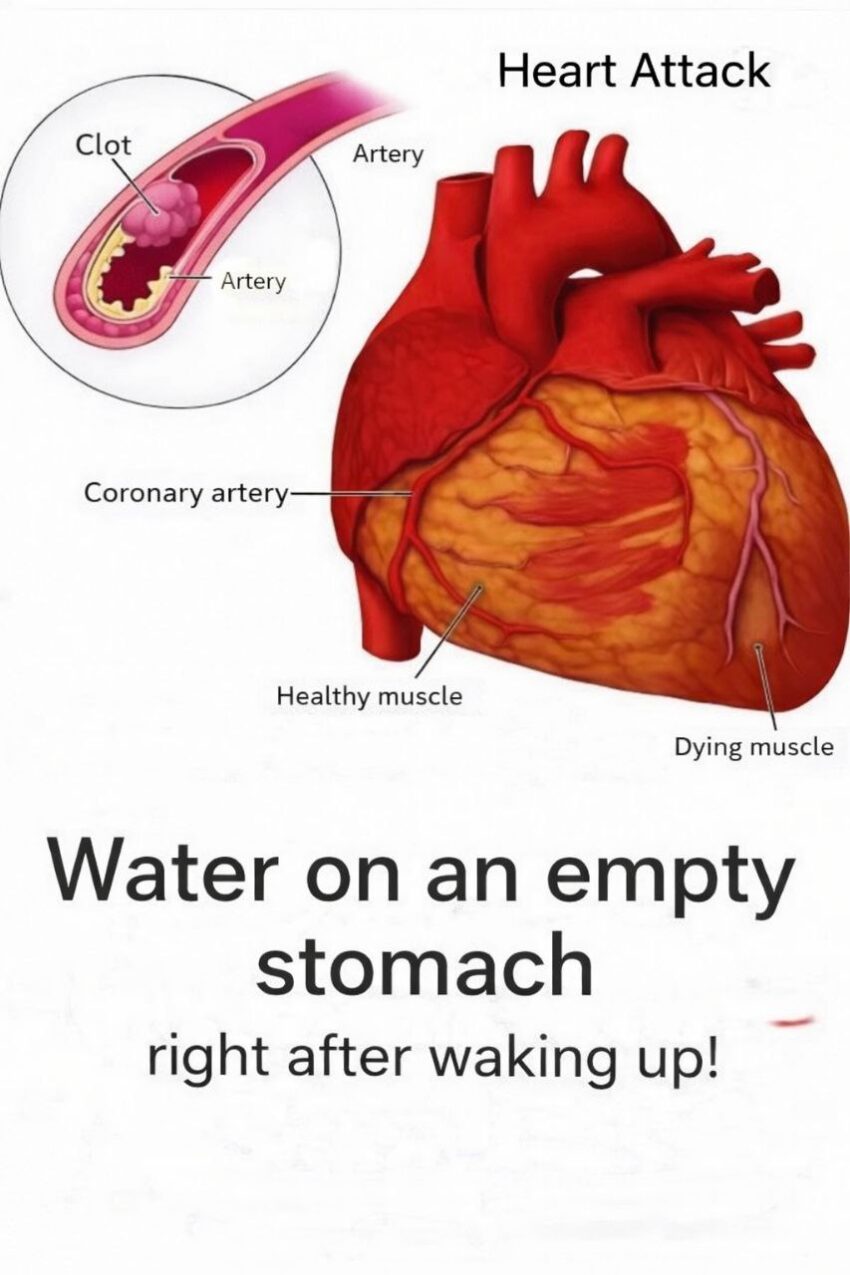
- Coronary arteries: Supply blood to the heart muscle itself.
- Pericardium: Protective outer sac of the heart.
🔹 Blood Flow Through the Heart
- Deoxygenated blood → RA → tricuspid valve → RV → pulmonary valve → pulmonary artery → lungs
- Oxygenated blood → pulmonary veins → LA → mitral valve → LV → aortic valve → body
💡 Tips for Reading a Heart Diagram
- Blue = deoxygenated blood, red = oxygenated blood.
- Arrows usually indicate blood flow direction.
- Valves prevent backflow and ensure unidirectional flow.
If you want, I can create a labeled, simplified heart anatomy diagram showing all chambers, valves, and major vessels for easier understanding.
Do you want me to do that?
