Here’s a detailed overview of the human gallbladder anatomy:
1. Location
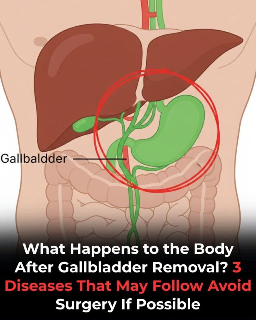
- The gallbladder is a small, pear-shaped organ located underneath the liver on the right side of the abdomen, in a depression called the gallbladder fossa.
- Lies posteroinferior to the right lobe of the liver.
- Typically measures 7–10 cm in length and 4 cm in diameter when fully distended.
2. Structure
The gallbladder has three main parts:
- Fundus
- Rounded end that projects beyond the lower border of the liver.
- Often palpable in thin individuals.
- Body
- Largest portion; lies against the visceral surface of the liver.
- Stores bile temporarily.
- Neck
- Narrow, tapered portion leading to the cystic duct.
- Contains Rokitansky-Aschoff sinuses, small mucosal outpouchings.
3. Internal Features
- Mucosa: Lined by simple columnar epithelium, specialized for absorption of water and electrolytes.
- Muscular layer: Smooth muscle fibers contract to expel bile into the cystic duct.
- Serosa: Connective tissue layer covering most of the gallbladder; part that contacts the liver lacks serosa.
4. Connections
- Cystic Duct: Connects the gallbladder neck to the common hepatic duct, forming the common bile duct.
- Blood Supply:
- Cystic artery, usually a branch of the right hepatic artery.
- Venous Drainage: Cystic veins drain directly into the liver (portal system).
- Innervation: Autonomic nervous system (sympathetic from celiac plexus, parasympathetic from vagus nerve).
5. Function
- Storage of bile: Produced by the liver.
- Concentration of bile: Absorbs water and electrolytes to make bile more concentrated.
- Release of bile: In response to cholecystokinin (CCK) after a fatty meal, the gallbladder contracts, sending bile through the cystic duct into the common bile duct and then to the duodenum.
6. Clinical Relevance
- Gallstones (cholelithiasis): Crystals forming in bile, often lodging in the gallbladder or cystic duct.
- Cholecystitis: Inflammation, usually due to gallstones or infection.
- Gallbladder removal (cholecystectomy): Common treatment for symptomatic gallstones.
If you want, I can also make a labeled diagram of the gallbladder showing its parts, ducts, and blood supply, which makes understanding its anatomy much easier.
Do you want me to make that diagram?
